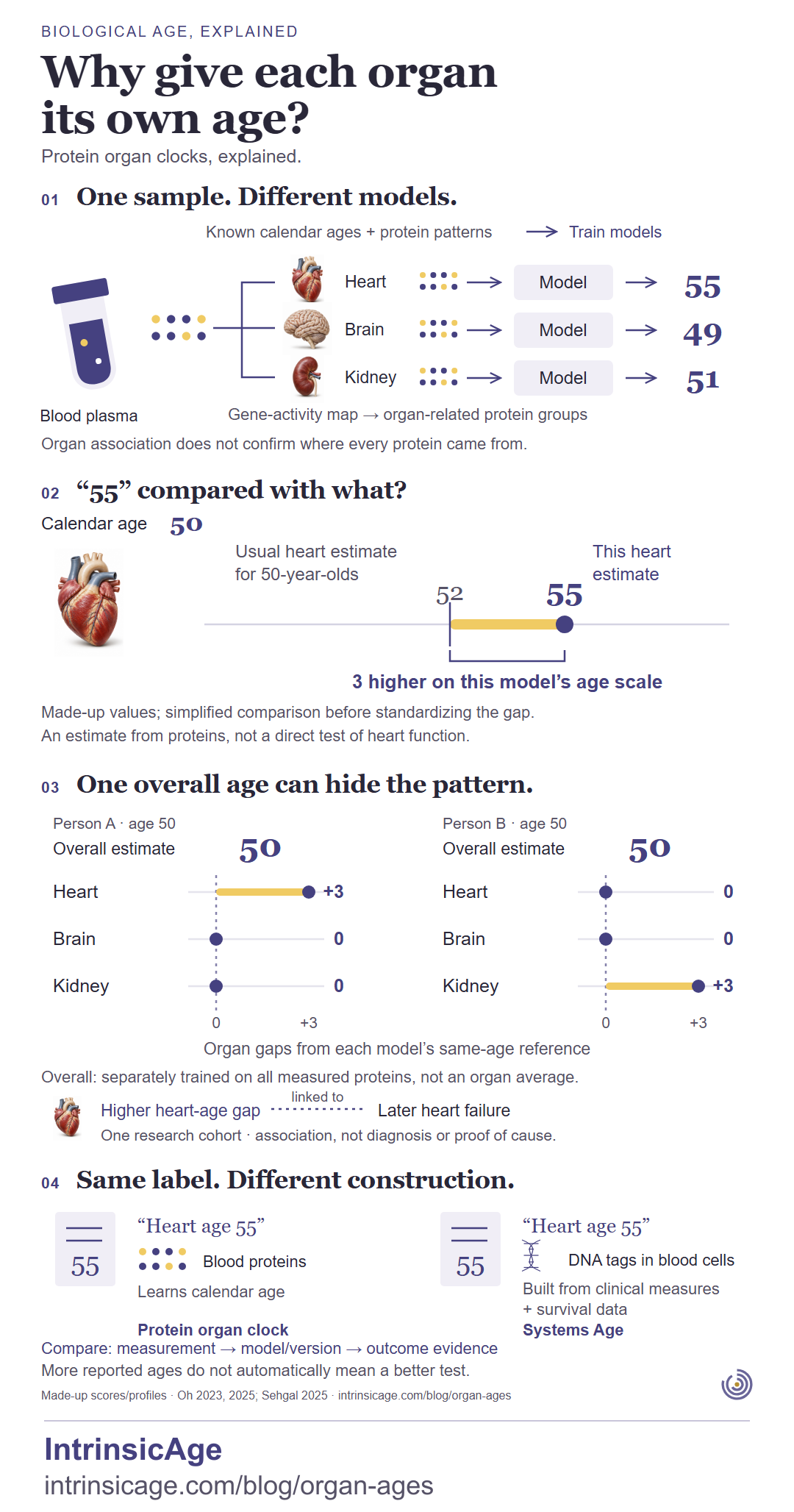
Known calendar ages and protein patterns train the models. A gene-activity map groups organ-related proteins measured in blood plasma. Separate heart, brain and kidney models produce example estimates of 55, 49 and 51. Organ association does not confirm where every protein came from.
55 compared with what?
For a person whose calendar age is 50, the model's usual heart estimate for 50-year-olds is 52. This person's heart estimate is 55: 3 higher on the model's age scale. These are made-up values and a simplified comparison before standardizing the gap. The estimate comes from proteins, not a direct test of heart function.
One overall age can hide the pattern.
Made-up profiles for two people aged 50; both have an overall estimate of 50. Organ gaps are compared with each model's same-age reference.
Organ
Person A
Person B
Heart
+3
0
Brain
0
0
Kidney
0
+3
The overall estimate is separately trained on all measured proteins, not an organ average. In one research cohort, higher heart-age gaps were linked to later heart failure: an association, not a diagnosis or proof of cause.
Same label. Different construction.
Two illustrative reports both say “heart age 55”, but their construction differs.
Two ways to construct an age estimate.
Method
Measurement
Construction
Protein organ clock
Blood proteins
Learns calendar age.
Systems Age
DNA tags in blood cells
Built from clinical measures and survival data.
Compare the measurement, model/version and outcome evidence. More reported ages do not automatically mean a better test.
Sources: Oh et al., Nature 2023 and Nature Medicine 2025; Sehgal et al., Nature Aging 2025. The article's references link to the papers.
How organ-age estimates are built, how to read their reference, and what separate organ profiles reveal. All displayed scores and profiles are made up.
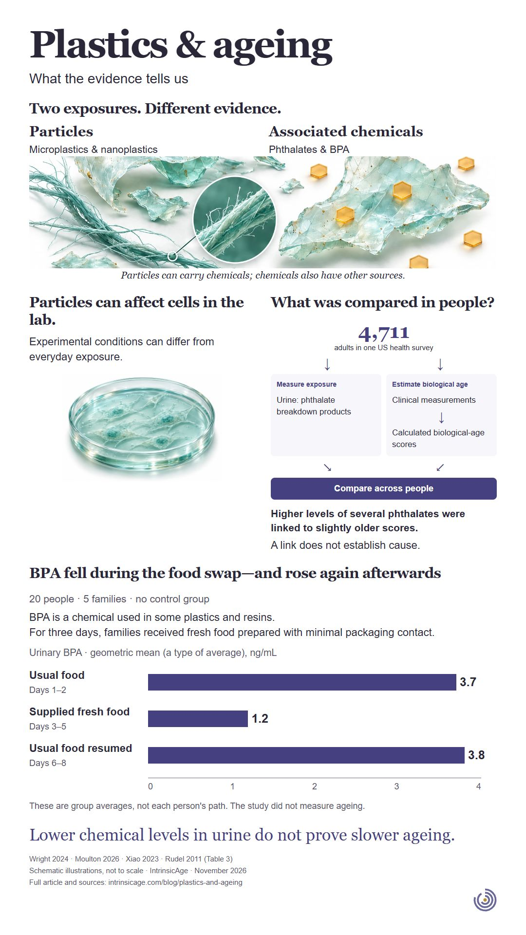 Plastics & ageingWhat the evidence tells usTwo exposures. Different evidence.ParticlesMicroplastics & nanoplasticsAssociated chemicalsPhthalates & BPA
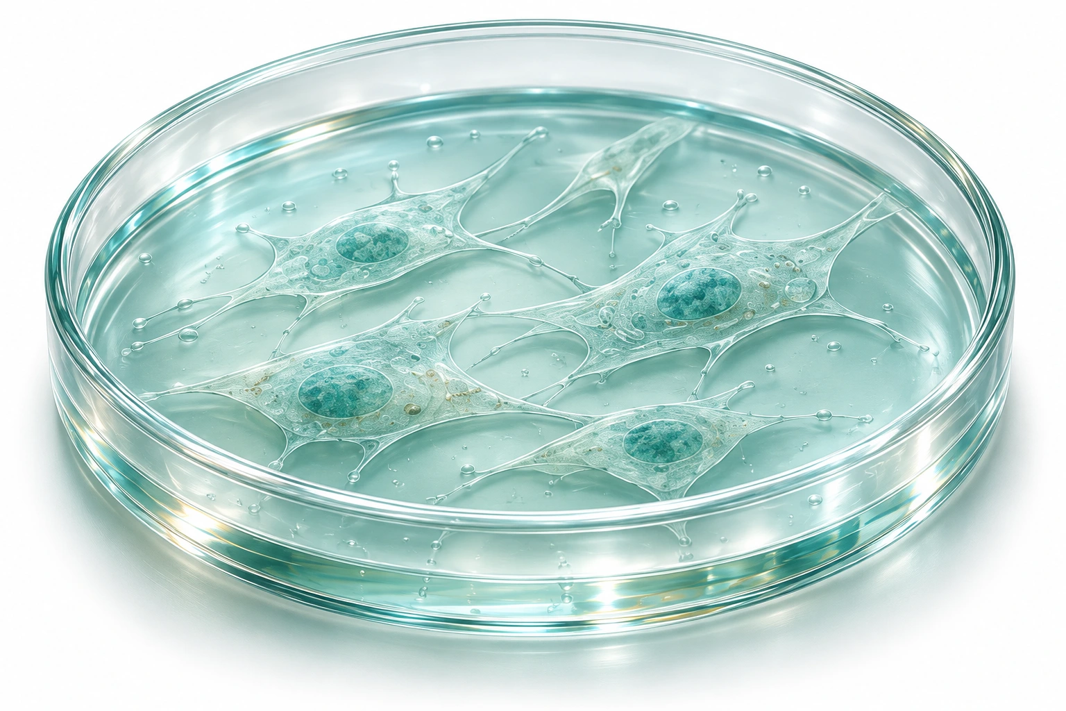
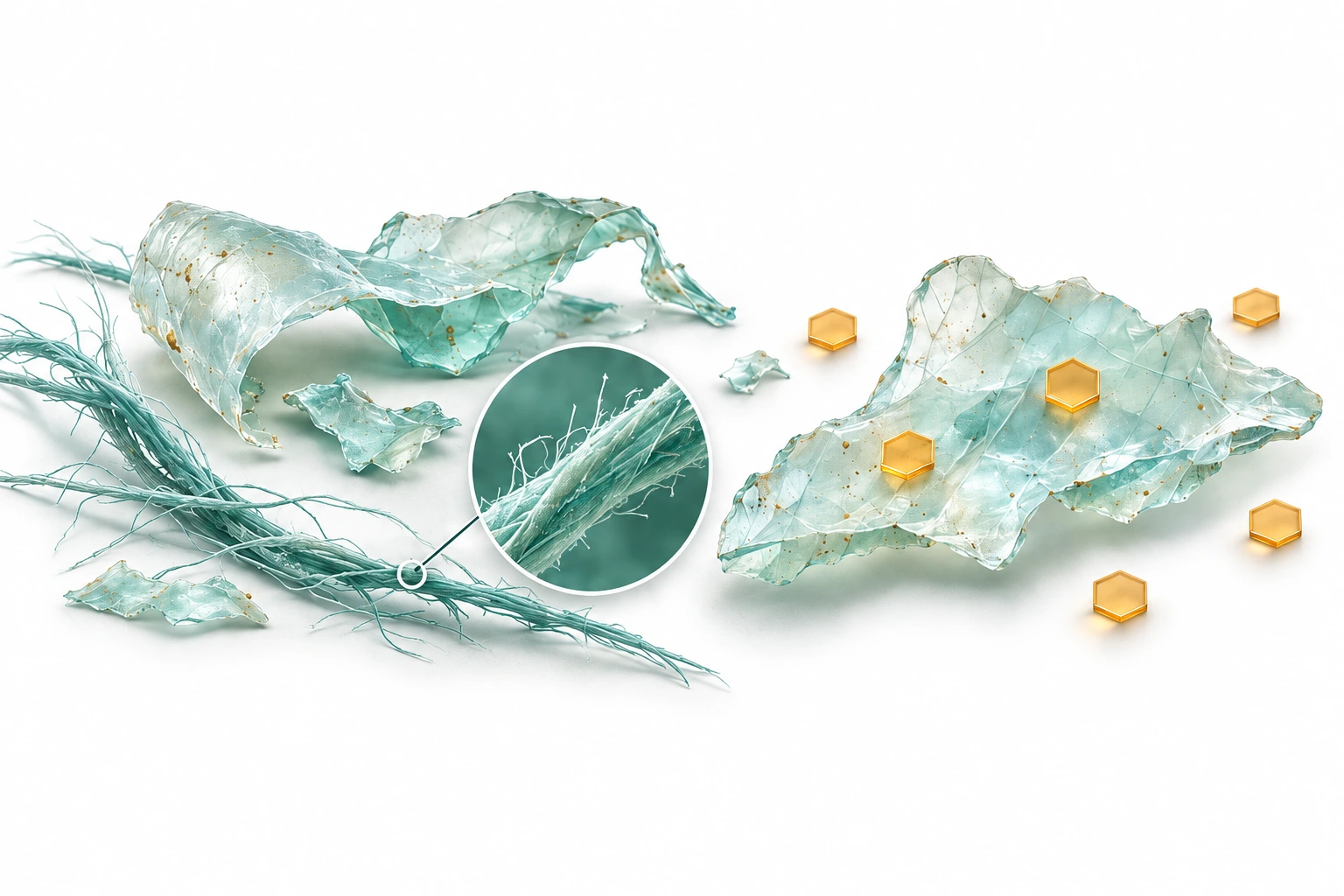 Particles can carry chemicals; chemicals also have other sources.Particles can affect cells in the lab.Experimental conditions can differ from everyday exposure.
 What was compared in people?4,711adults in one US health surveyMeasure exposureUrine: phthalate breakdown productsEstimate biological ageClinical measurementsCalculated biological-age scoresCompare across peopleHigher levels of several phthalates were linked to slightly older scores.A link does not establish cause.BPA fell during the food swap—and rose again afterwards20 people · 5 families · no control groupBPA is a chemical used in some plastics and resins.For three days, families received fresh food prepared with minimal packaging contact.Urinary BPA · geometric mean (a type of average), ng/mLThese are group averages, not each person's path. The study did not measure ageing.Lower chemical levels in urine do not prove slower ageing.Wright 2024 · Moulton 2026 · Xiao 2023 · Rudel 2011 (Table 3)